Kimura Group
 Research Associate TAKEO, Yoko
Research Subjects
- Development of high-precision X-ray optical devices using ultra-precision fabrication and measurement techniques
- Development of new X-ray microscopy technology using advanced light sources
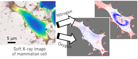
- Material property imaging with soft X-ray microscopy
- Femtosecond imaging of samples in liquids using X-ray free-electron lasers
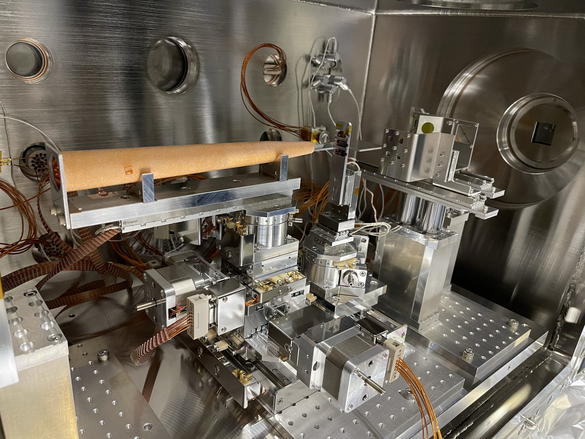
Our laboratory is engaged in the development of next-generation X-ray imaging technologies that combine state-of-the-art X-ray sources—such as X-ray free-electron lasers, synchrotron radiation, and high-order harmonics—with ultra-precise X-ray optical components. Our research includes the construction of advanced X-ray microscopes at large-scale synchrotoron radiation facilities such as SPring-8 and SACLA, the design and fabrication of X-ray optics using cutting-edge semiconductor manufacturing techniques with atomic-level precision, and the development of sophisticated computational algorithms for lensless imaging. In close collaboration with domestic and international research partners, we actively apply these technologies to a wide range of samples, including inorganic materials such as nanoparticles and magnetic nanostructures, as well as biological specimens like mammalian cells and marine plankton. By uncovering the relationship between mesoscopic structures and their physical or chemical properties with high spatial and temporal resolution, we aim to open up new frontiers in a variety of scientific fields, including materials science, nanotechnology, and life science.