
Time-Resolved X-Ray Photoelectron Diffraction Using an Angle-Resolved Time-of-Flight Electron Analyzer
PI of Joint-use project: K. HayashiHost lab: I. Matsuda Group
Photoelectron spectroscopy (PES) is a powerful experimental approach for investigating chemical composition, electronic states, and atomic structure of a material. Recently, the PES measurement method is conducted with temporal resolutions and the experiment becomes a successful method to track these quantities during the dynamical events of a sample. At SPring-8 BL07LSU, such a time-resolved PES technique is developed and the beamline endstation has been opened for users. In the present research, we combined it with angular resolution and demonstrated a time-resolved experiment of X-ray photoelectron diffraction (XPD) [1].
The time-resolved measurements were performed by a pump-probe method that combined synchrotron radiation (SPring-8 BL07LSU) and laser (BL07LASER) pulses [2,3]. Photon energy of synchrotron radiation was set at hv = 300eV, while that of laser at hv = 1.55 eV. Angle-resolved spectra were obtained with a two-dimensionally angle-resolved time-of-flight (2DARTOF) analyzer [3]. The photoelectrons, detected by the 2D delay line detector, were recorded at the arrival time (t) and position (x, y), subsequently converted into energy (E) and 2D emission angle (θx, θy). A demonstration experiment was made on a silicene layer, a honeycomb lattice of Si atoms, on a ZrB2 substrate. Figures 1 (a) and 1 (b) show the Si 2p XPD patterns of two atomic sites, α and β, in the silicene. The two sites appear at different binding energies of PES, as shown in Fig. 1 (c). One can find in Fig.1 (a,b) that the experimental XPD pattern is reproduced by the simulation. Focusing on the pattern at the β site, the temporal variation can be detected after the delay time. As presented in Fig. 1(d), the features, labeled A and B, shows temporal changes, weakening, disappearance, and recovery, during sub-nanoseconds (ns) to 10 ns. These results indicate that the time-resolved XPD experiment can be effectively made with a 2DARTOF analyzer. The novel approach is useful to examine the structural changes during dynamical events, such as photocatalysis and photo-induced phase transition.
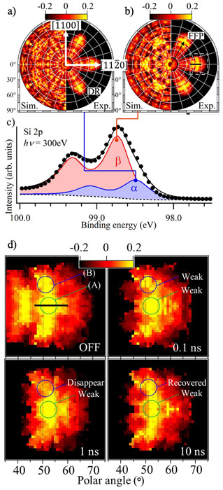
Fig. 1. (a,b) XPD patterns of the Si 2p core levels at the α and β sites in (c) XPS spectrum. (d) Time-resolved XPS pattern of the β site taken at various delay times [1].
References
- [1] A. K. Ang, Y. Fukatsu, K. Kimura, Y. Yamamoto, T. Yonezawa, H. Nitta, A. Fleurence, S. Yamamoto, I. Matsuda, Y. Yamada-Takamura, and K. Hayashi, Jpn. J. Appl. Phys. 59, 100902 (2020).
- [2] J. Miyawaki, S. Yamamoto, Y. Hirata, M. Horio, Y. Harada, and I. Matsuda, AAPPS Bulletin 31, 25 (2021).
- [3] M. Ogawa et al., Rev. Sci. Instrum. 83, 023109 (2012).