
Chiral Spin Spiral Structure Studied by Spin-Polarized Scanning Tunneling Microscopy
Hasegawa Group
Because of the potential application for future spintronics devices, spiral magnetic structures, such as skyrmion lattices, domain walls, and homogeneous spin spiral structures, have been a subject of extensive studies. Various types of spin rotations have been reported; Bloch-type (helical) or Néel-type (cycloidal), left-handed (↑←↓) or right-handed (↑→↓), and chiral or non-chiral. To determine these types, neutron scattering, Lorentz microscopy, etc. have been utilized. For nanometer- or atomic-scale spin spiral structures, often formed in ultrathin films, spin-polarized scanning tunneling microscopy (SP-STM), which detects spin orientations in atomic-scale spatial resolution, is the only method for the determination. We have investigated the types of the spin structure observed in a monolayer (ML) Mn thin film formed on a tungsten substrate using the ultimate spin-probe microscopic method.
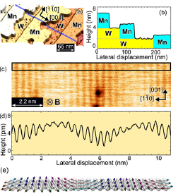
Fig. 1. (a) STM image of a Mn monolayer (ML) formed on a W(110) substrate. (b) Cross-sectional profile taken along the blue line in (a). (c) Spin-polarized STM image of 1 ML Mn/W(110) taken with an Fe-coated W tip magnetized perpendicular to the sample surface. (d) Cross-sectional profile averaged in the boxed area in (c). (e) schematic of the magnetic structure of ML Mn/W(110).
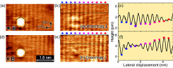
Fig. 2. (a, d) SP-STM images of 1 ML Mn/W(110) taken in the same area with an Fe-coated W tip magnetized in the in-plane (a) and out-of-plane directions (d) The direction of the applied magnetic field is shown by the arrows. (b, e) Images whose nonmagnetic contribution was subtracted from (a) and (d). (c, f) Cross-sectional profiles averaged in the boxed areas of (b, e). The same colored dots indicate the same atomic rows in the images of (b, c, e, f).
In magnetic systems whose inversion symmetry is broken, the Dzyaloshinskii–Moriya interaction (DMI) plays a key role in the formation of chiral spin structures. Since the symmetry is naturally broken at interfaces, several systems composed of 3d magnetic thin films and 5d non-magnetic heavy-elemental substrates showing a strong spin-orbit coupling exhibit DMI-driven chiral spin structures. For the investigation of how overlayers and substrates affect the polarity and strength of DMI a W(110) substrate is one of the ideal systems because various chiral structures have been discovered on it with overlayers. From SP-STM images taken with a tip whose magnetization direction is well controlled, we revealed that the Mn monolayer indeed exhibits a cycloidal spin spiral structure with a left-handed rotation (See a schematic shown in Fig. 1(e)). By comparing with e.g. the case of magnetic domain walls observed in Fe thin films, we found that the polarity of DMI is basically determined by the substrate.
The SP-STM experiments were performed with an ultrahigh vacuum STM at 5 K using Fe-coated W tips as a magnetic probe. Since the amount of the tunneling current is proportional to the cosine of the angle θ between the tip and sample magnetization directions, one can obtain the spin or magnetic contrast of the sample using the magnetized tip. A two-axis superconducting magnet was used to align the tip magnetization to the specific orientations of the sample.
A typical STM image taken on monolayer Mn-covered W(110) surface is shown in Fig. 1(a). A cross-sectional profile shown in Fig. 1(b) indicates atomically flat terraces of Mn ML grown from the W step edges. Figure 1(c) shows an SP-STM image taken on an ML region of Mn/W(110) with a tip magnetized perpendicular to the sample surface. Bright and dark rows separated by an atomic distance along the [110] direction indicate antiferromagnetic magnetization of the Mn [001] rows. The contrast vanishes periodically at the Mn rows whose magnetization directions are close to the in-plane direction (i.e., θ is close to 90°), indicating a spin-spiral structure.
There are two possible types of spin spiral structure to explain the observed images; a cycloidal structure whose spins are rotating in the (001) plane and a helical one rotating in the (110) plane. To determine the rotational type of the spin structure, SP-STM images are taken with a tip magnetized along the [001] direction. The obtained images showed suppressed magnetic contrast, indicating a cycloidal spin spiral structure. This result is consistent with the mechanism of the interfacial DMI, which only produces cycloidal spin spiral structures or Néel-type domain walls.
Then, in order to clarify the rotational sense of the cycloidal spin spiral structure, we performed SP-STM measurements with a spin-polarized tip magnetized in two orthogonal directions within the (001) plane. When we focus on every two adjacent spins, the magnetic contrast exhibits a sinusoidal variation. By changing the direction of the tip magnetization from parallel to perpendicular to the surface [i.e. from the leftward to the upward direction], we should observe +90° or -90° phase shifts in the sinusoidal profile depending on the rotational sense.
Figures 2(a, d) show SP-STM images taken in the same area with the tips magnetized in the directions shown in the figures. White clusters provide a proof that it is the same area. In order to extract the magnetic contrast, we subtracted the nonmagnetic contributions, which are derived by averaging two SP-STM images taken with the tips magnetized in opposite directions. Cross-sectional profiles shown in Figs. 2(c, f) indicate the direction of the phase shift is consistent with that expected for the left-handed spin spiral structure.
The domain walls in the Fe DL/W(110) were found as Néel-type domain walls with right-handed rotation, which can be explained by the consequence of the competition between ferromagnetic interaction (↑↑) and right-handed DMI (↑→). On the other hand, the left-handed rotation of Mn ML (↑↘←↗↓) is a consequence of the competition between antiferromagnetic interaction (↑↓) and right-handed DMI (↑→). We thus found the consistent role of the substrate on the polarity of DMI in the both cases.
References
- [1] M. Haze, Y. Yoshida, and Y. Hasegawa, arXiv:1604.01123 (2016).