
A Compact Permanent-Magnet System for Measuring Magnetic Circular Dichroism in Resonant Inelastic Soft X-Ray Scattering
J. Miyawaki, S. Suga, and Y. Harada
Resonant inelastic X-ray scattering (RIXS), especially soft X-ray RIXS (SX-RIXS), has made remarkable progress in energy resolution over the past few decades in conjunction with the improvement of the performance of synchrotron radiation sources and the advancements in the RIXS instrumentation. Accordingly, RIXS has come to occupy an important position in X-ray spectroscopy techniques for investigating the bulk electronic structures. The photon-in/photon-out process adds to RIXS the distinctive feature that the electronic structure can be measured even in an electronic and/or magnetic field. Regarding the use of the magnetic field for RIXS, magnetic circular dichroism (MCD) in RIXS (RIXS-MCD) has intensively been used to extract spin-resolved valence band excitations. In addition to all the features inherited from RIXS itself, RIXS-MCD of dd excitations that can easily be separated by the use of the latest high-energy-resolution RIXS apparatus can provide magnetic information for each localized d orbital. With these advantages, RIXS-MCD is becoming a very promising technique, and is expected to be successfully applied to new functional materials in which spins play important roles, such as multiferroics and those to be used for spintronic devices. In order to demonstrate the significance of RIXS-MCD with high energy resolution and encourage its wider use, it is highly desired that RIXS-MCD experiments can be performed as a common tool at any RIXS end-station in worldwide synchrotron radiation facilities. Therefore, we developed a compact and portable magnet system for RIXS-MCD composed of Nd-Fe-B permanent magnets which can generate a magnetic field of ~0.25 T [1].
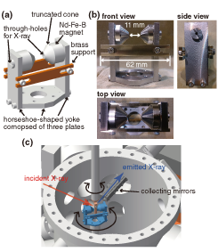
Fig. 1. Schematic representation and photographs of the magnet system and experimental setup. (a) Schematic representation and (b) photographs of the magnetic circuit. The magnets are fixed by a screw with a through-hole for the X-ray beam. (c) Experimental configuration of RIXS-MCD at the HORNET end-station in SPring-8 BL07LSU when the angle of the magnetic field is set at 45° to the incident X-ray.
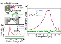
Fig. 2. Fe L-edge XMCD and RIXS-MCD spectra of an α-Fe2O3(111) single crystal. (a) Sketch of the experimental configuration with the orientation of the spin. The magnetic field is parallel to the incident X-ray and the incident angle is 10° from the sample surface. Yellow arrows on the α-Fe2O3(111) sample represent the direction of the antiferromagnetically coupled and canted spin moments on the (111) plane. (b) Fe L2,3-edge XAS spectra measured by circularly polarized X-rays with the XMCD spectrum. (c) RIXS-MCD spectra measured at hv = 713.25 eV indicated by a vertical line in (b).
Figures 1(a) and 1(b) display a schematic drawing and photographs of the originally-designed magnetic circuit. The magnetic circuit consists of Nd-Fe-B magnets, a horseshoe-shaped yoke, truncated cone poles, and two supports. The Nd-Fe-B magnets are the source of the magnetic field. The horseshoe-shaped yoke and truncated cone poles are made of Fe, and functions to focus the magnetic fields. The two supports are made of brass, and function to resist the attractive force between the magnets. The overall size of the magnetic circuit is within φ65 mm so that it can be installed through and mounted on the most commonly used 4-1/2 inch-diameter ConFlat® flange. This compact design of the magnetic circuit allows it to be installed without any modification to the existing vacuum chambers and optical systems for RIXS. Figure 1(c) is a schematic representation of the experimental configuration in a vacuum chamber using this magnet system at the HORNET end-station in SPring-8 BL07LSU.
We have demonstrated the capability of the present magnet system for RIXS-MCD. Figure 2 shows X-ray magnetic circular dichroism (XMCD) and RIXS-MCD of weak ferromagnetism in α-Fe2O3. We measured XMCD and RIXS-MCD in the grazing incidence geometry as shown in Fig. 2(a). No discernible signal was detected in its XMCD spectrum [Fig. 2(b)]. This is because the net magnetic moment owing to the weak ferromagnetism is too small to be detected. Figure 2(c) exhibits the RIXS-MCD spectra excited at the charge-transfer satellite as indicated by the solid line in Fig. 2(b). RIXS-MCD was clearly observed at 1.8 and 4.85 eV loss energies, which are assigned to a dd excitation and a charge-transfer transition, respectively. Thus, RIXS-MCD was clearly observed by high-energy-resolution RIXS, though XMCD was not observed. These results confirmed that the observed RIXS-MCD was induced by the RIXS process, via the different relaxation probabilities of polarized valence electrons to polarized core holes created by the circularly polarized X-rays. These results suggest that more useful bulk information about the ground-state spin state can be obtained by using RIXS-MCD.
References
- [1] J. Miyawaki, S. Suga, H. Fujiwara, H. Niwa, H. Kiuchi, and Y. Harada, J. Synchrotron Rad. 24, 449 (2017).