
Membrane Tubulation and Polyhedral Vesicle Formation Induced by Banana-Shaped Proteins
Noguchi Group
In living cells, membrane morphology is regulated by various proteins. Many membrane reshaping proteins contain a Bin/Amphiphysin/Rvs (BAR) domain, which consists of a banana-shaped rod. The BAR domain bends the biomembrane along the rod axis and the features of this anisotropic bending have recently been studied. We study as to how such a local anisotropic curvature induces effective interaction between proteins and changes the global shape of vesicles and membrane tubes using meshless membrane simulations. The proteins are modeled as banana-shaped rods strongly adhered to the membrane.
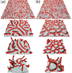
Fig. 1. Sequential snapshots of tabulation from a flat membrane induced by protein rods. (a) Positive perpendicular rod curvature. (b) Negative perpendicular rod curvature.
Our study revealed that the rods assemble via two continuous directional phase separations in membrane tubes and vesicles unlike a conventional two-dimensional phase separation [1]. As the rod curvature increases, the rods assemble along the azimuthal direction and subsequently along the longitudinal direction accompanied by shape transformation of the membrane tube. In the vesicle, in addition to these two assembly processes, further increase in the rod curvature induces tubular scaffold formation. We also found that the polyhedral vesicles and polygonal tubes are stabilized at high rod densities [2]. The discrete shape transition between triangular and buckled discoidal tubes and between polyhedral shapes are obtained. As line tension of the membrane edge is reduced, the protein adhesion induces membrane rupture leading to high-genus vesicle formation and vesicle inversion [3]. These shape transformations and assemblies are not obtained by isotropic inclusions.
We also studied the effects of the spontaneous (side) curvature perpendicular to the rod, which can be generated by protein–protein and membrane–protein interactions [4]. We revealed that the perpendicular curvature can drastically alter the tubulation dynamics from a flat membrane at high protein density whereas no significant difference is obtained at low density. A percolated network is intermediately formed depending on the perpendicular curvature. This network suppresses tubule protrusion. Thus, this kinetic trap makes tubule formation significantly slower. The stability of network structures can be explained by a simple geometric model. Positive surface tensions and vesicle membrane curvature can stabilize this network structure by suppressing the tubulation. It is known that tubulation dynamics can be different even for proteins consisting of the same BAR domains. Our finding suggests that the interactions between the rest parts of the proteins can give significant effects.
References
- [1] H. Noguchi, EPL 108, 48001 (2014).
- [2] H. Noguchi, J. Chem. Phys. 143, 243109 (2015).
- [3] H. Noguchi, Phys. Rev. E to be published (arXiv:1602.04569).
- [4] H. Noguchi, Sci. Rep. 6, 20935 (2016).